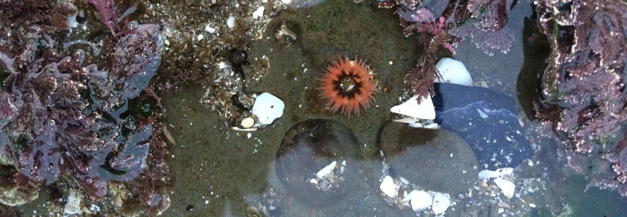
One of the most common questions I get when teaching my students about urchins is: “Can they move?” The answer is: yes!
Urchins move around by utilizing tube feet powered by their water vascular system–a system that uses water pressure to extend and retract the tube feet. Sea stars more famously utilize a similar system to control their tube feet. The first step of the water vascular system is the madreporite–in this case, a large, porous plate at the top of the urchin.

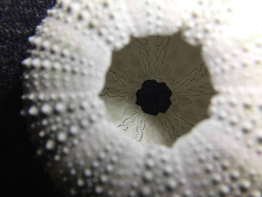
It’s a little tough to see on this specimen, but there are ten apical plates surrounding that center hole in the test. Five apical plates have an eyespot and five have an gonopore for releasing gametes. One of those gonopore plates also holds a madreporite.
The madreporite is typically covered in skin while the urchin is alive and is rarely still present on dead urchin tests. The apical plates often break off from the urchin test and are easily lost. I was thrilled to find that this urchin test still had its madreporite!
Once water is brought in through the madreporite, it passes through a series of canals before reaching the tube feet. Tube feet are connected to ampullae, which help the tube feet extend and retract.
Imagine an eyedropper filled with water connected to a thin plastic bag. Squeezing the eyedropper pushes the water out into the plastic bag, extending it. Sucking the water back into the eyedropper retracts the plastic bag. That’s pretty much how tube feet operate! The ampullae are the eyedroppers and the plastic bags are the tube feet.
Here’s a video of tube feet in action that I took under a microscope:
If you look closely, you can actually see particles moving down on the left and up on the right in the closer tube foot. Those are coelomocytes–cells present in the water vascular systems of urchins. They mark the direction of fluid travel on each side of a dividing septum. An extended tube foot has constant flow to keep it extended.
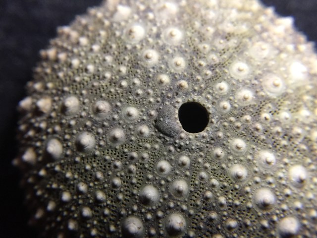
You can actually see exactly where tube feet extend from by looking at the inside of an urchin test.
Those five groups of holes in wavy lines are the holes where tube feet poke out. In life, the ampullae would be lining those holes on the inside, each one controlling a tube foot. The groups of tube feet and groups of large spines alternate on urchin tests. If you look closely, you can actually see those holes on the outside of the urchin test too.
And those are the basics of urchin tube feet! Next urchin post: how and what urchins eat!

Hey, just went to your Davidson class, here’s how to remove that “BLOG AT WORDPRESS.COM.” footer (not written by me): https://www.wpbeginner.com/wp-themes/how-to-remove-the-powered-by-wordpress-footer-links/
LikeLike