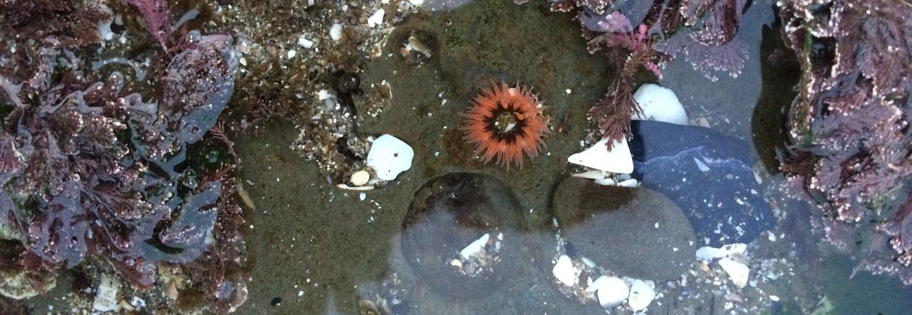
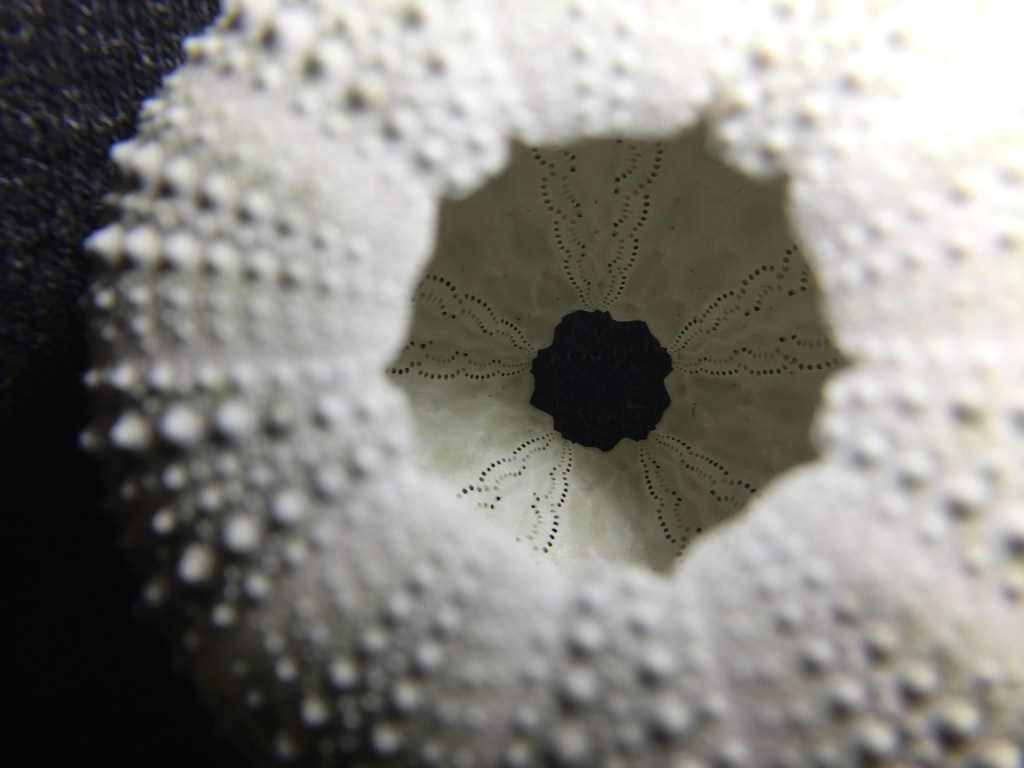
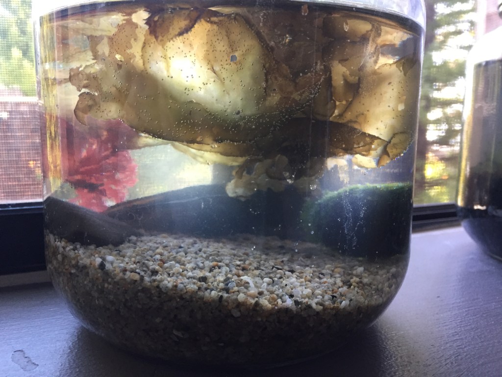
The Uniqueness of Urchins: Spines

Sea urchins–purple urchins especially–have a bit of a bad reputation on the West Coast of the United States right now. They are currently forming and maintaining urchin barrens, which are kelp forests’ “evil twin” or alternative stable state. But that’s a complicated story for another time!

Even though urchins aren’t exactly universally loved, I am fond of them. It probably has something to do with my general love for marine invertebrates. Regardless, there’s lots to learn about urchins! Let me show you with the help of some specimens, starting with their spines.

This is one of my specimens–a dried purple sea urchin with its spines still intact. As you can see, several of its spines have broken off over time. They can move each spine individually to defend against attacks. If you “tickle” an urchin on its exoskeleton, its spines will respond in a matter of seconds.

For reference, here’s a smaller, living purple sea urchin:

The color has faded in the dead specimen–living urchins are a brighter purple.

Typically, dead urchins don’t retain their spines for long. The spines tend to fall off, along with the skin, revealing what’s known as the test. The test is the urchin’s main, circular exoskeleton.

A purple urchin test

The spines have sockets that connect to the ball-like bumps protruding from the test. This enables them to rotate each spine. Unlike arthropods, urchins grow their test and spines continuously–no need to molt!

Urchins exhibit pentaradial symmetry, which means that their bodies are radially symmetrical in multiples of five. Sea stars and other echinoderms are also pentaradial. This test shows how urchins have alternating stripes of spine-bump sizes.

The little holes in the test are for the tube feet to extend from. You’ll hear more about tube feet in a future blog post!

It’s important to mention that some urchins have venomous spines–particularly tropical species. Neither purple urchins nor the closely related red urchins have venom in their spines, so they are completely safe to touch. The only threat these temperate urchins pose to humans is the threat of getting spines stuck in your skin.

Urchins can drop spines when living for a variety of reasons, including disease, duress, or trying to dispose of a fouling organism. It’s possible for urchins to recover from dropping a few or many spines if given enough recovery time, especially if it has enough nutrients in the water and in its diet.

Those are the basics about urchin spines! Stay tuned for more urchin facts and pictures!

Investigating kelp in Minecraft (Activity for Middle School and Junior High students)

Since I teach online, I’m always coming up with new activities for my students to complete. I thought I would share one of the activities in my Underwater Forests: Kelp Ecosystems course I’m teaching this semester. We’re looking more closely at Macrocystis pyrifera this week, so I’m assigning a Minecraft activity where students will observe and experiment with Minecraft’s version of kelp. They are not only learning more about real life giant kelp, but they’re also practicing running experiments.

For my students who follow me here–spoilers!

Minecraft recently added kelp into the game, which gives us the perfect opportunity to investigate its properties!

Find a natural kelp forest in Minecraft. 

Plant a kelp individual in fairly deep water. How long does it take to grow a block higher? Time it! Try it again with a few more individuals. Is the time the same for all of them?

Plant a kelp individual in shallow water. How long does it take for it to grow a block higher? Time this one too and try it with several individuals! Does it take a shorter amount of time?

Compare this data to real kelp organisms. Do you think the time it takes for kelp to grow would differ between shallow and deep water? If so, how?

Finally, take a look at the surface of the water. Does the kelp keep growing at the surface? How does this compare to a real kelp forest?

Write down your results and thoughts in at least six interesting sentences. Good luck & have fun!

I’m looking forward to seeing what they discover!

Marine Miniature Ecosystem(s) Update #2 (A fast-paced update!)

Quite a lot has happened to the jar ecosystems since I last wrote. For one, the first ecosystem did not go as well as hoped.

As it turned out, there were a few more polychaetes than expected…as in, approximately five. I discovered them shortly after posting the first update.

There’s one now!

There were also two larger roundworms and several smaller worms that were extremely difficult to photograph!

One of the larger roundworms hanging out at the surface of the water.

Although I’d hoped to return the polychates to the ocean in the morning, there was unfortunately not enough dissolved oxygen for them to survive the night. After they died, the nutrient influx was massive and caused the ecosystem to collapse.

Some lovely nutrient sludge draped over rotting algae.

I still had another jar to fill with seawater, but I waited. My Marine Ecology course was starting in only a few days and I wanted my students to be involved in the jar creation process. In the best case scenario, our class jar would illustrate how ecosystems function and ecosystem processes. In the worst case scenario, it would illustrate how ecosystems are very difficult to get right and how we can learn from every experiment, regardless of whether it turned out correctly.

I polled the class and they decided to create the jar as follows:

  • With both coarse and fine sand
  • Water from “As far out into the ocean as Emma can reasonably wade”
  • Green intertidal algae and red intertidal algae
  • Mussel shell and a small rock as decorations

After a quick trip to the ocean, our jar was created!

A fresh start.

After a week and a half of slow deterioration, the class decided to add more algae and remake my first jar attempt with the same characteristics as before.

The algae in the class jar was looking a little worse for wear, but there were some signs of life–like this roundworm.

Back to the ocean I went. Once I returned, I rearranged algae and tried to aerate the jars a bit to get more dissolved oxygen in the water. My dorm room had definitely smelled better! I attempted to capture as much diversity as possible, since diversity generally increases ecosystem stability.

Old (2nd attempt) jar on the left, new jar on the right.

In exciting news for the class, our new jar had unknowingly brought home a new class mascot: a little nudibranch!

The class voted to name them “Peanut butter.”

Unfortunately, after a couple days, Peanut butter disappeared. We came to terms with the loss as a class with a moment of silence.

The original jar wasn’t doing well either, even with the addition of more algae. However, this was a great example of alternative ecosystem states for the class to learn from! It can be very difficult to push a jar ecosystem back into a different ecosystem state.

Fast forward a few weeks and both jars had seen better days.

(The jars were also moved home, since the quarter ended!)

Before moving back home for winter break, I noticed that there seemed to be a growing layer of white around the circumference of the jar. My students and I were very interested in what this could be, so I took a sample to school and took a look under the microscope.

Bacterial mat?
Lovely!

The consensus was bacteria, especially since I also saw paramecia (which feed on bacteria). There might be hope for life in these jars!

Today, the jars are doing about the same.

A picture after they’ve been stirred up a bit.

I’m looking forward to watching them as they progress in these alternative stable states!

Marine Miniature Ecosystem Update #1

I realized a dream of mine today– a dream to create my very own miniature marine ecosystem. As a marine ecology student, the idea immediately appealed to me.

I’d been researching the concept for months and finally had the materials to put it into action! So I drove up to Davenport (where it’s legal to collect algae) this morning and created my brand-new ocean-in-a-jar.

From this angle, you can see Ulva and a red algae I have yet to positively ID. It’s a little beaten up and might actually be two species growing together.

The red algae has some interesting animal life of its own. It has some sort of bryozoan species encrusting on it, which I’m still in the process of identifying. It also has a few tiny ostrich plume hydroids. I’m interested to see if they have survived/will survive. Although I was sad to take them from their habitat, I rescued this algae and its epibionts from the wrack line. If the bryozoans and hydroids aren’t already dead, they would’ve been shortly thereafter.

Here you can see the (empty) mussel shell with encrusting coralline algae.

I included this mussel shell and another mussel half-shell in an attempt to provide algae a place to settle and provide shelter for organisms.

Here you can see the salt residue on the side of the jar and some Cladophora columbiana.

The algae has already begun photosynthesizing, which I was extremely happy to see! You can see some of the air bubbles still attached to the Cladophora.

Earlier today, there was an interesting development.

The unmistakable bristled sides of a polychaete worm!

It seems that a stowaway came along! I rinsed off all the algae in the ocean, but it seems that this one is good at holding on.

Polychaetes are a type of annelid (segmented) worms. The term polychaete means many bristles, which are attached to the little extensions (parapodia) along its body. This particular polychaete uses its bristles and parapodia to “swim” through the water.

I decided that it was a little big to be in the jar so back to the ocean we went. Although it seems that this is some sort of clam worm, which does eat algae, it seemed like too much to have in a jar that was just starting out.

Ready for release!

The release went well and the little polychaete scurried away from the sunlight fairly quickly. When I returned, I was shocked to find another surprise waiting for me!

Another one?! (it’s hanging onto the red algae)

Apparently, one of these types of algae is a common hiding place for these polychaetes! Since this one is a little smaller, I’m going to see how it does in the jar for now. I can’t find it right now, so hopefully it’s found a good place to live.

I’m looking forward to watching the jar change and finding out what other organisms appear!

If you are interested in creating your own ecosystem, I highly recommend checking out the YouTube channel Life in Jars?, which offers tutorials, information, and entertaining jar updates:

https://www.youtube.com/channel/UC0XNssyypOLiq4vVgXm9NtQ

Stay tuned for updates on this jar and on the next ecosystem I create…

Investigating my first specimen: Pyrosoma atlanticum

In the summer of 2018, I had the opportunity to participate in a biological and anthropological field study to the Channel Islands of California, specifically Santa Cruz Island. The trip was headed (in part) by my zoology professor from my community college, who assisted me in several marine identification efforts.

Of course, I was most looking forward to the rocky intertidal exploration. We all woke up bright and early at about five in the morning and headed out to the rocky intertidal zone. There were several highlights, including the first octopus I’d ever seen in the wild, a chiton that refused to release itself from the bowl it’d been placed in, a sea hare, and the awesome visible radula of a wavy top turban snail. However, perhaps the most exciting part of the trip was lying on the beach on our walk back. We found this organism:

While holding our new, accidentally-collected pet chiton in a bowl, my zoology professor and I attempted to identify what in the world this could be. It looked a little like a sponge, but there was no point of possible attachment. There was only one opening– the other end of the creature was sealed shut.

Obviously, there was only one solution. We put it in a water dish and I balanced two dishes of water on the (extremely bumpy) trip back to the campsite.

When we returned, we put both the mysterious, unidentified specimen and the chiton into the refrigerator while we packed up. I hypothesized that the chiton would loosen its grip if cooled down, so into the refrigerator it went. We decided to pack the specimen in the cooler in a plastic bag so we could further investigate when we returned to the research station, since we needed to keep to our schedule. Meanwhile, we had successfully cooled Titan the Chiton down enough that he detached from the bowl and were now pursuing the side mission: Release Titan the Chiton.

Later that day, after Titan the Chiton had been successfully released, the specimen was retrieved and we were ready for investigation!

It smelled pretty disgusting. However, that provided a valuable hint! It smelled like rotting animal flesh, not rotting algae, so algae was ruled out.

It was also spewing slimy goo, which was certainly an interesting experience. After about an hour of attempting to identify it, we had no luck. But that was fine with me, because that meant…

…I got to take it home for further investigation! At dinner, we discussed the unknown nature of the specimen. One of my classmates immediately pulled out her phone and took to Google. After only a couple of searches, she actually found a picture of it! We learned that it was Pyrosoma atlanticum, which is a type of tunicate colony.

Tunicates, members of Urochordata, are closely related to vertebrates and thus humans. Adult tunicates, like the ones making up this colony, only have two out of the five chordate features: pharyngeal gill slits and the endostyle. They filter feed using mucus created by the endostyle, which coats the gill slits, which was likely the mucus it was spewing. There are both solitary and colonial tunicates– colonial tunicates like this one connect zooids (individual tunicates) through a common “tunic.”

Thankfully, we also learned it was definitely dead by the time we got it, because it wasn’t producing any blue light through bioluminescence.

Once we returned home and slept for a couple days, I brought my prized specimen to the zoology lab, where we could actually take a look with a dissecting microscope. After carefully slicing it, this is what we found:

Here, you can see the excurrent siphons pointing inward (the small pyramid-like structures on the bottom surface). These siphons propel the colony through the water. Each zooid has its own excurrent siphon.

My very first specimen now sits on my desk at UCSC in a place of honor.